« Back to Glossary IndexStructure and Function of the Frontal Lobe:
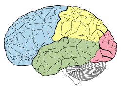
– The frontal lobe is the largest lobe of the brain.
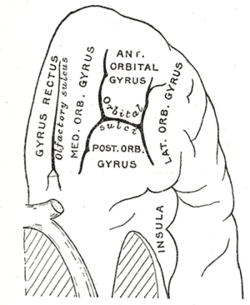
– It is divided into lateral, polar, orbital, and medial parts.
– Gyri in the frontal lobe include superior frontal, middle frontal, and inferior frontal gyri.
– The frontal cortex is considered the action cortex.
– The prefrontal cortex is responsible for reasoning and projecting future consequences of actions.
– It integrates non-task based memories.
– Controls voluntary movement, decision making, and language.
Clinical Significance of Frontal Lobe Damage:
– Damage can result from strokes, mini-strokes, or traumatic brain injuries.
– Symptoms include inappropriate responses, lack of emotional expression, and loss of motivation.
– Can lead to personality changes and decreased executive functions.
– Confabulation and uncharacteristic cheerfulness are less common effects of damage.
– Specific syndromes like delusional misidentification and Capgras syndrome can occur.
Genetic and Neurological Influences on the Frontal Lobe:
– DNA damage increases with age, affecting genes crucial for learning and memory.
– Prenatal alcohol exposure can lead to damage.
– Variants in genes like COMT can impact performance in working memory tasks or increase schizophrenia risk.
– Reduced expression of key genes in the frontal cortex occurs with aging, affecting synaptic plasticity and mitochondrial function.
Psychosurgical Procedures and Historical Context:
– Psychosurgery in the early 20th century involved damaging pathways connecting the frontal lobe to the limbic system.
– Frontal lobotomy was a historical procedure that reduced distress but had severe side effects.
– Modern procedures like anterior capsulotomy and bilateral cingulotomy are more precise and used for obsessional disorders and depression.
– Frontal lobotomy had a high mortality rate, leading to its decline.
Research and Comparative Studies on the Frontal Lobe:
– Theories of frontal lobe function are categorized into single-process, multi-process, construct-led, and single-symptom theories.
– Ongoing research aims to develop a unified theory of frontal lobe function.
– Studies challenge the view of human frontal lobe enlargement compared to other primates.
– Human cognition is attributed to greater connectedness rather than cortical volume.
– Comparative studies with great apes show similarities in frontal cortex size.
Frontal lobe (Wikipedia)
The frontal lobe is the largest of the four major lobes of the brain in mammals, and is located at the front of each cerebral hemisphere (in front of the parietal lobe and the temporal lobe). It is parted from the parietal lobe by a groove between tissues called the central sulcus and from the temporal lobe by a deeper groove called the lateral sulcus (Sylvian fissure). The most anterior rounded part of the frontal lobe (though not well-defined) is known as the frontal pole, one of the three poles of the cerebrum.
The frontal lobe is covered by the frontal cortex. The frontal cortex includes the premotor cortex and the primary motor cortex – parts of the motor cortex. The front part of the frontal cortex is covered by the prefrontal cortex. The nonprimary motor cortex is a functionally defined portion of the frontal lobe.
There are four principal gyri in the frontal lobe. The precentral gyrus is directly anterior to the central sulcus, running parallel to it and contains the primary motor cortex, which controls voluntary movements of specific body parts. Three horizontally arranged subsections of the frontal gyrus are the superior frontal gyrus, the middle frontal gyrus, and the inferior frontal gyrus. The inferior frontal gyrus is divided into three parts – the orbital part, the triangular part and the opercular part.
The frontal lobe contains most of the dopaminergic neurons in the cerebral cortex. The dopaminergic pathways are associated with reward, attention, short-term memory tasks, planning, and motivation. Dopamine tends to limit and select sensory information coming from the thalamus to the forebrain.
 Marko Kadunc


